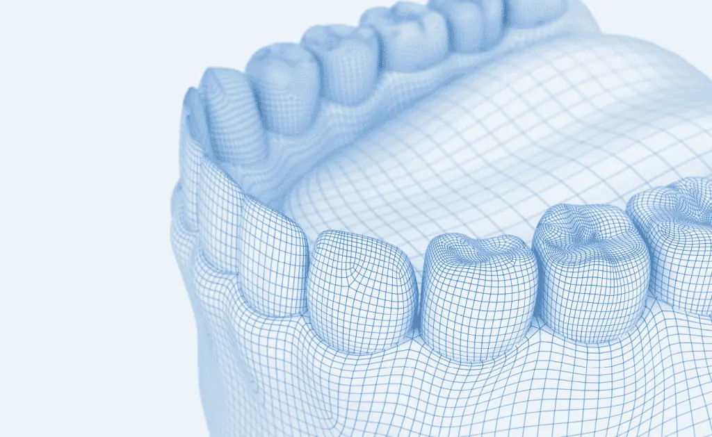
Thanks to the latest technology, we now have the opportunity to take three-dimensional images of the mouth area of our patients using digital volume tomography (DVT).
More insight, less radiation exposure
The special feature of the 3D X-ray procedure is that the finest detail is visible thanks to the perfect image quality, whereas we would not usually be able to see this. 3D technology offers new perspectives and insights into the oral region. And even better: there is very low exposure to radiation; indeed, this is 80% lower than with CT.
Advantages of the 3D X-ray procedure
- Low radiation exposure
- The X-ray process takes place seated or standing
- Clear presentation thanks to 3D images
- Precise diagnosis and treatment planning
- Minimises the risks of an operation
- Fast and straightforward archiving of X-ray documents
- Saves time as you do not have to attend a radiology practice or hospital for the CT
Zahnfleischbluten bei der täglichen Zahnpflege sowie Rötung und Schwellung des Zahnfleisches können die ersten Anzeichen für eine Parodontitis sein. Gehen Sie dann am besten sofort zum Zahnarzt. Durch die Früherkennung haben Sie die besten Chancen auf Heilung.
3D X-ray - the safety advantage
3D technology offers us a completely new dimension in terms of safe diagnosis. We can use it to pre-plan surgical and implant procedures. 3D X-ray procedures are the ideal examination method in many other areas too, for example, for:
Diagnosis
3D X-rays are the perfect aid for the initial and checking examinations. Sites of inflammation can also be located reliably. Wisdom teeth and other displaced teeth can be identified precisely in this way.
Planning and checking for teeth replacements
Using the 3D X-ray procedure, we can assess your teeth, bones and root fillings much more precisely and thereby make a more exact long-term diagnosis for your teeth.
Root fillings
We can recognise and treat inflamed root tips and special features of the root canal system in a targeted way prior to the treatment using 3D technology.
Gum disease
We can use this procedure to identify how the bones around the tooth roots have degraded, and plan the treatment accordingly.
Surgical procedures
The nerves, sinus cavity and structures of the bone can be looked at in the finest detail thanks to the 3D X-ray process. For operation procedures such as putting in implants or removing wisdom teeth, this is an invaluable safety advantage.
Procedures and examinations in the jaw area
For orthodontic planning in particular, a 3-dimensional examination is an advantage. Planned measures for patients can be coordinated very effectively. We can achieve better results in jaw joint diagnoses and functional diagnostics using this technique.
Sie haben Fragen rund um das Thema 3-D-Röntgen?
You will find answers to the most frequently asked questions here.
So-called digital volume tomography (DVT) is used with the 3D X-ray procedure. This prepares X-ray images - as you will know from traditional computed tomography. The special feature of DVT is that it moves right around the jaw area and therefore captures a great many individual images. The computer calculates a three-dimensional, high-resolution image of the skull from these individual images. The dentist sees the pathway of the jaw and facial nerves, the situational relationships of the teeth, the composition of the bone as well as dental and jaw diseases.
Yes, that's right. The radiation exposure is eighty per cent lower than with traditional computer tomography. This is due to the fact that the DVT generates X-ray images with an extremely short length of exposure. The radiation dose is therefore much lower.
On the one hand, much lower radiation exposure, and on the other hand, the perfect image quality. You can see even the most minute detail on the 3D images. This enables precise diagnosis and treatment planning.