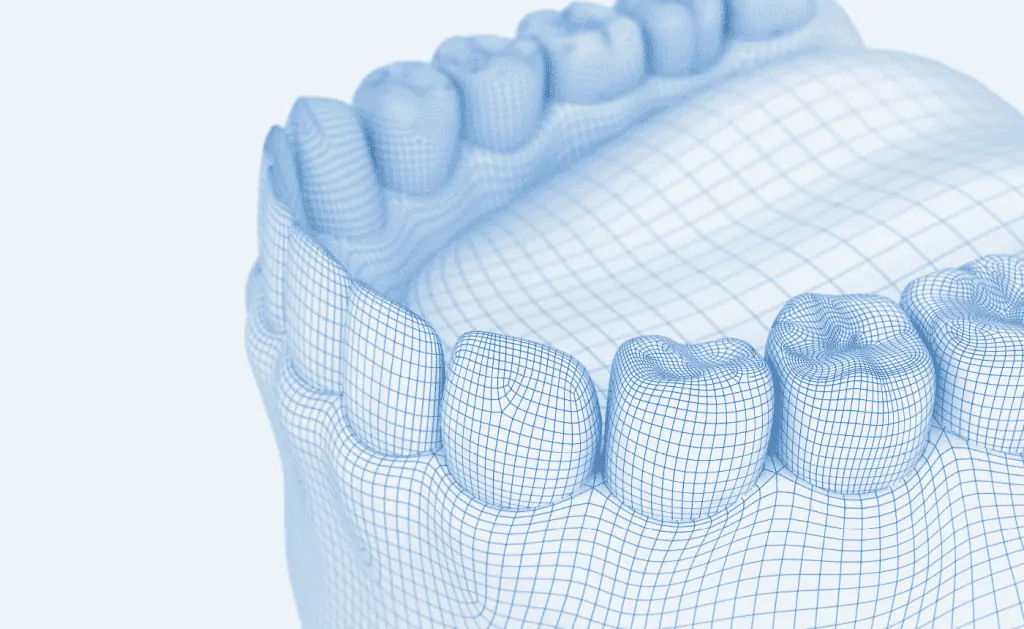
Thanks to state-of-the-art technology, we now have the ability to create three-dimensional images of our patients’ oral areas using digital volume tomography (DVT).
Greater insight, lower radiation exposure
The unique feature of 3D X-ray technology is that its perfect image quality reveals the finest details that would otherwise remain hidden. 3D technology offers new perspectives and insights into the oral region. Even better: radiation exposure is very low—80% less than with a CT scan.
Advantages of 3D X-ray technology
- Low radiation exposure
- X-ray procedure performed while sitting or standing
- Clear visualization through 3D images
- Precise diagnosis and treatment planning
- Minimization of surgical risks
- Fast and straightforward archiving of X-ray documents
- Time savings, as you do not need to visit a radiology practice or hospital for a CT scan.
Not every dental issue requires a 3D X-ray. We use digital volume tomography selectively and only when it offers genuine diagnostic value—such as for reliable planning, precise diagnoses, and minimally invasive procedures.
3D X-rays – an extra level of safety
3D technology offers us a whole new dimension of diagnostic reliability. It allows us to plan surgical and implantological procedures more accurately. 3D X-ray technology is also the ideal examination method in many other areas, such as:
Diagnosis
3D X-rays are the ideal tool for initial and follow-up examinations. Foci of inflammation can also be reliably detected. Wisdom teeth and other impacted teeth can be precisely localized.
Planning and monitoring of dental prosthetics
With 3D X-ray technology, we can assess your teeth, bone, and root fillings much more precisely, allowing for a more accurate long-term prognosis for your teeth.
Root canal fillings
Using 3D technology, we can identify inflamed root tips and specific characteristics of the root canal system before treatment and provide targeted care.
Gum disease
This procedure allows us to clearly see the extent of bone loss around the tooth roots and plan treatment accordingly.
Surgical procedures
Thanks to 3D X-ray technology, we can see nerves, maxillary sinuses, and bone structures in minute detail. This provides an invaluable increase in safety during surgical procedures, such as placing implants or removing wisdom teeth.
Procedures and examinations in the jaw area
Three-dimensional examination is particularly advantageous for orthodontic planning. This allows planned measures to be even better tailored to the patient. We can also achieve better results in temporomandibular joint and functional diagnostics.
Do you have questions about 3D X-rays?
Here you will find answers to the most frequently asked questions.
3D X-ray technology utilizes what is known as a Digital Volume Tomograph (DVT). It produces X-ray images similar to those from conventional computer tomographs. The unique feature of DVT is that it rotates around the entire jaw area, taking numerous individual images. These images are then used by a computer to calculate a three-dimensional, high-resolution image of the skull. This allows the dentist to see the path of jaw and facial nerves, the spatial relationship between teeth, bone quality, and any dental or jaw diseases.
Yes, that is correct. Radiation exposure is 80% lower than with conventional computer tomographs. This is because the DVT generates X-ray images with an extremely short exposure time, resulting in a much lower radiation dose.
Firstly, the significantly lower radiation exposure, and secondly, the perfect image quality. Even the smallest details are visible on 3D images, enabling precise diagnosis and treatment planning.